
Radiology plays a central role in today’s healthcare by enabling detailed internal assessments without the need for invasive procedures. Women’s imaging, as a specialised branch, focuses on female-specific health conditions, offering accurate diagnostics and care solutions across all life stages.
The use of imaging technologies has revolutionised how health conditions are identified, monitored, and treated. By providing a clear view of internal organs, bones, and soft tissues, radiology supports early diagnosis and guides treatment decisions with greater precision. From chronic illness management to emergency assessments, it helps medical teams respond quickly and effectively.
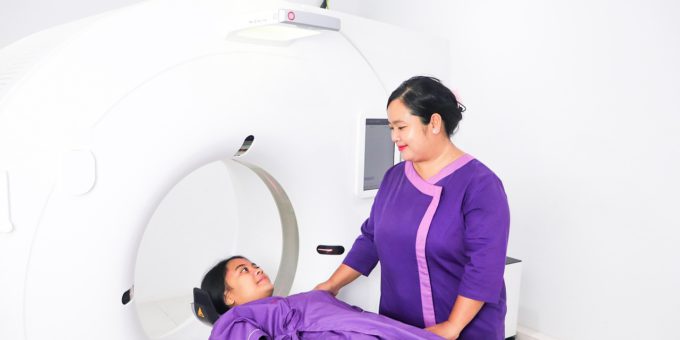
Medical imaging encompasses a variety of techniques. Each type serves a specific purpose and delivers unique insights. X-rays are often used to view bones and detect fractures, while ultrasound is widely applied to monitor internal organs and observe pregnancies. MRI and CT scans allow for high-resolution views of soft tissues, helping to detect issues like tumours, inflammation, or vascular irregularities. These tools reduce uncertainty in diagnosis and support timely interventions.
Women’s imaging offers specialised assessments aimed at addressing health issues such as breast conditions, pelvic disorders, and pregnancy-related concerns. Regular mammograms, pelvic ultrasounds, and prenatal scans help detect changes early, often before symptoms arise. These tools are essential in promoting preventive care and improving long-term outcomes for women across all age groups.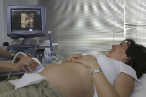
Ultrasound remains one of the most common imaging options in women’s health. Its ability to visualise reproductive organs, monitor fetal development, and assist with fertility evaluations makes it an essential part of routine care. Since it does not involve radiation, it is particularly safe for expectant mothers. Beyond pregnancy, ultrasound is also used to detect fibroids, cysts, and irregularities in the uterus or ovaries.
Screening mammography continues to be a standard approach to breast cancer detection. When performed regularly, it helps identify abnormalities at a stage when treatment is most effective. For individuals with dense breast tissue or increased risk, additional imaging like breast MRI or 3D mammography may be recommended to provide a clearer picture.
Bone health is another critical area where imaging offers valuable insights. As the risk of osteoporosis rises with age, particularly in postmenopausal women, bone density scans (DEXA) help evaluate fracture risk and inform treatment strategies. These scans are simple, quick, and effective for the early detection of bone loss.
Technological advancements are reshaping how images are captured, analysed, and shared. Artificial intelligence is being integrated into diagnostic tools to support radiologists in detecting patterns and anomalies. By enhancing image interpretation, AI can improve accuracy and reduce the time between scanning and diagnosis.
Cloud-based image sharing is also streamlining access. Patients and healthcare providers can now view and transfer imaging results more efficiently through digital platforms. This improvement reduces delays, eliminates the need for physical films, and supports collaboration among specialists regardless of location.
Radiologists are highly trained medical professionals who play a vital role behind the scenes. They interpret medical images, collaborate with referring doctors, and provide expert opinions that guide treatment. Their work is not limited to reading images; it also involves understanding patient history, recommending appropriate imaging, and ensuring that procedures are safe and appropriate for each individual.
Patient-centred care is becoming increasingly important in imaging services. Efforts are being made to create a more welcoming environment through clear communication, shorter waiting times, and better support during procedures. Women undergoing imaging—particularly those for sensitive or anxiety-inducing conditions—often benefit from facilities designed with privacy and comfort in mind.
Raising awareness about the benefits of routine imaging is another key aspect of improving health outcomes. Many individuals delay or avoid scans due to fear, misunderstanding, or lack of information. Educating patients on what to expect and how imaging supports their health can encourage more timely and consistent participation in recommended screenings.
Personalised medicine is another area where radiology plays a growing role. Imaging results contribute to customised treatment strategies that consider individual differences in anatomy, disease progression, and risk factors. This tailored approach helps ensure that care is as effective and efficient as possible.
Accessibility to imaging services continues to improve with the introduction of mobile units and remote radiology consultations. These solutions are especially valuable in rural and regional areas where medical resources may be limited. Through tele-radiology, medical images can be reviewed by specialists remotely, ensuring that patients receive timely and expert evaluations regardless of their location.
Ongoing research continues to push the boundaries of what imaging can do. Techniques are being developed to capture even more detailed images with lower doses of radiation. Others focus on real-time imaging during procedures, helping guide surgeries or biopsies with extreme precision. These developments offer exciting potential for faster diagnoses and improved treatment planning in the years ahead.
Ultimately, imaging is much more than a diagnostic tool—it is a critical component of proactive and preventive healthcare. Whether it’s through regular screenings, emergency evaluations, or monitoring treatment progress, radiology offers a window into the body that helps medical professionals make informed decisions. With its growing capabilities, it will continue to shape how health conditions are understood and managed in a wide range of clinical settings.